 KOR
KOR ENG
ENG





Home / 응용분야 / 상세페이지


Robust FFPE Nuclei Quantification Using EVE™ HT FL
The global demand for formalin-fixed paraffin-embedded (FFPE) tissue analysis continues to expand. Driven by advancements in precision medicine, these specimens have become increasingly important for biomarker discovery and targeted therapy development. Recently, FFPE tissues have been successfully integrated into advanced genomic workflows, such as single-nucleus RNA sequencing (snRNA-seq). Prior to snRNA-seq, precise nuclear counting is required to maximize target discovery rates while minimizing doublets.
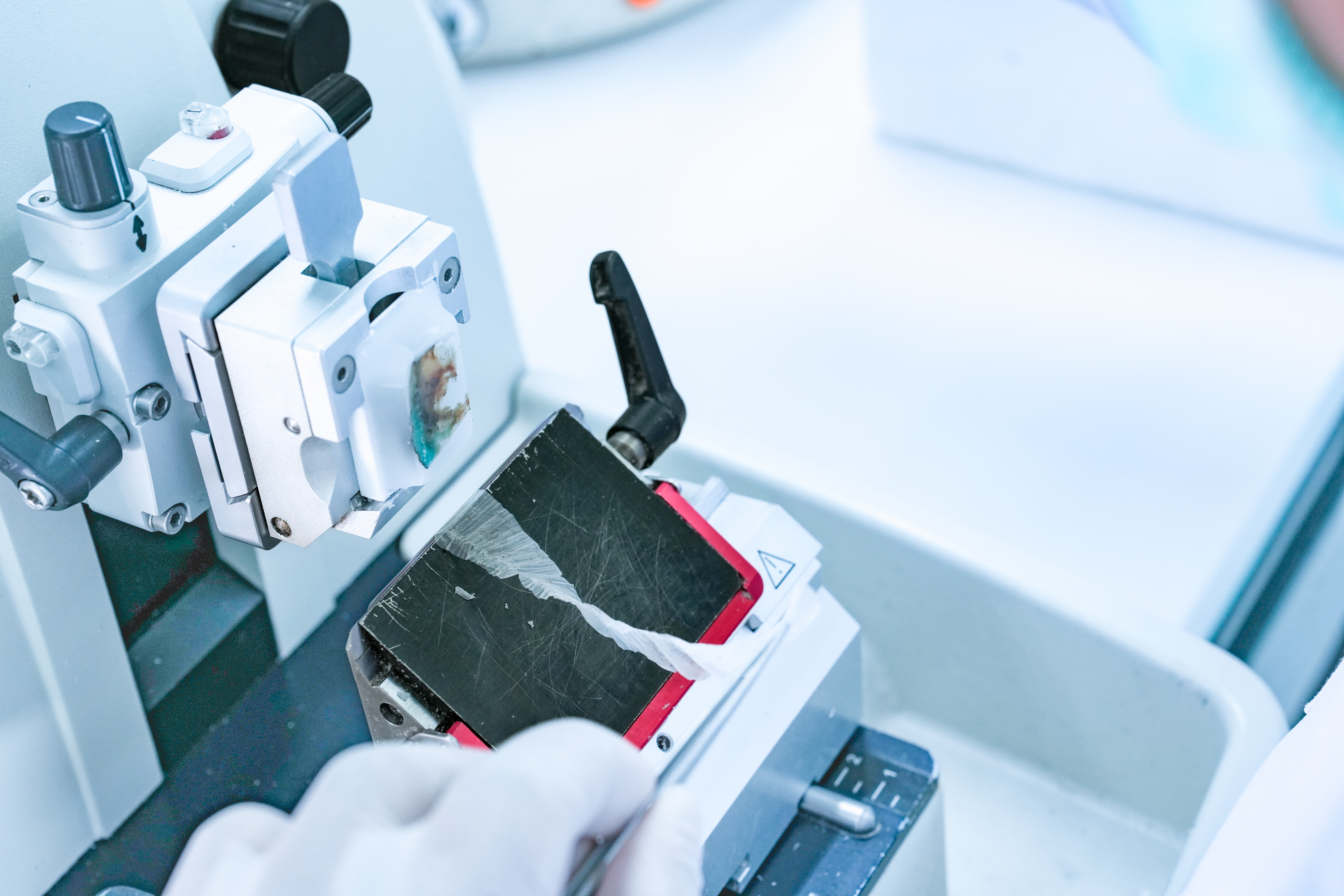
To meet the growing global demand for formalin-fixed paraffin-embedded tissue analysis, the EVE HT FL offers a dedicated FFPE mode.
1. Materials
• Mouse FFPE tissue sample
• Human FFPE tissue sample
• Xylene
• Ethanol (100%)
• Nuclease Free Water
• PBS
• Collagenase I & II (Roche, 5401135001)
• Proteinase K (Roche, 311586001)
• Mortar and pestle
• Cell strainer (porosity 30μm)
• Quenching buffer (10xGenomics, 1000414)
Staining dye & Instrument
• AO/DAPI with EVE™ HT FL
• YOYO-1 with EVE™ HT FL
• PI (N solution) with ADAM™ MC2
2. Methods
Deparaffinization and Rehydration of FFPE tissues
1. Wash twice in xylene for 10 min each.
2. Wash twice in 100% ethanol for 5 min each.
3. Wash once in 70% ethanol for 3 min.
4. Wash once in 50% ethanol for 3 min.
5. Wash once in nuclease-free water for 3 min.
6. Wash once in cold PBS for 5 min.
Tissue Dissociation and Nuclei isolation*
1. Suspend deparaffinized and rehydrated tissues in an enzyme solution like collagenase. Optionally, one can use proteinase K for hard-to-dissociate tissues.
2. Mechanically dissociate tissues in enzyme solution using a pestle.
3. Pass everything through a 30 μm filter.
4. Centrifuge at 700 RCF for 5 min.
5. Resuspend pellet in 1x quenching buffer.
6. Centrifuge at 700 RCF for 5 min.
7. Resuspend pellet in PBS.
8. Run cell counting.
* Nuclei isolation protocol can vary depending on tissue types and institutions. Users are expected to use a protocol that produces nuclei with little clumps or debris.
3. Results
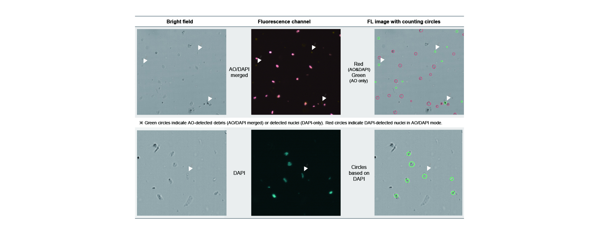
We first tried to stain nuclei isolated from FFPE tissues (Mouse) with EVE HT FL reagent (AO/DAPI). While mammalian cells stained with EVE HT FL reagents were bright in both AO and DAPI channels, FFPE nuclei stained with EVE HT FL reagents were very dim in both fluorescence channels. After increasing exposure time significantly, we were able to make them bright, and we found that nuclei were bright enough to be counted with EVE HT FL.
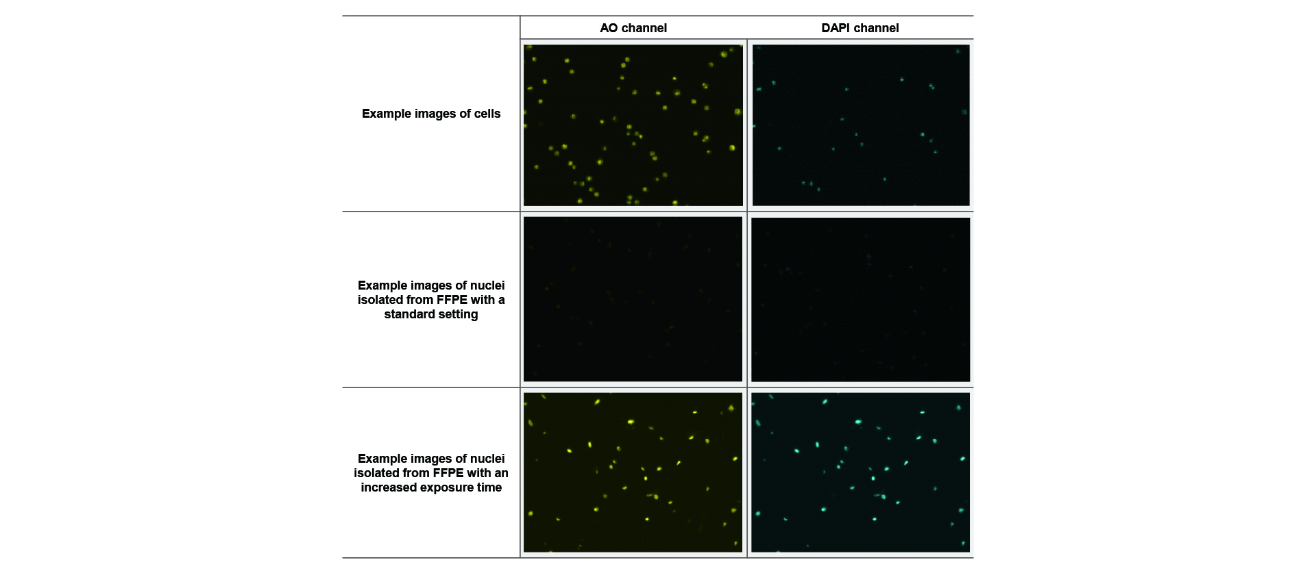
When we looked at images closely, we found that AO stained not only nuclei but also debris that did not look like cell nuclei. However, DAPI seemed to stain almost all nuclei, and EVE HT FL software could count nuclei with various shapes including elongated ones as shown in the example below.
4. Adding a new FFPE mode
Because AO has high probability to stain not only nuclei but also some of cell debris, counting based on AO images could be misleading. On the other hand, DAPI was found to have little tendency to stain non-nucleus debris. Therefore, we developed a new FFPE mode in which only brightfield and DAPI channels will be taken, and nuclei counts will be measured solely from DAPI.

5. Comparison with other staining methods
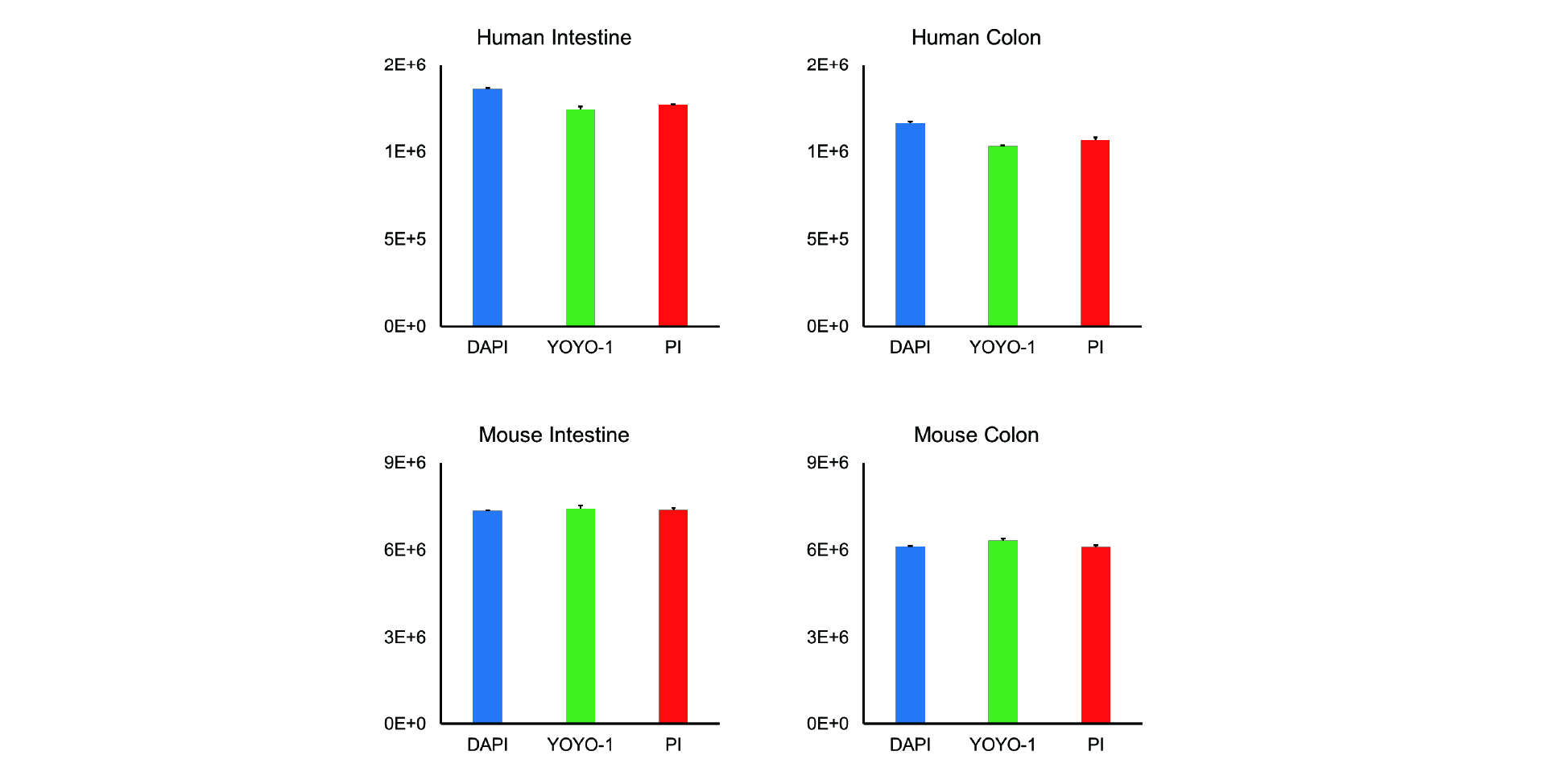
NanoEntek’s single slide fluorescence cell counter, ADAM-MC2 uses PI (propidium iodide) to count nucleated cells. Because PI is known to be nuclei-specific, we compared PI and DAPI with nuclei isolated from FFPE tissues. In addition to them, we have also tried YOYO-1. We have tested these 3 dyes with various FFPE tissues. All the results suggest that DAPI on EVE HT FL and PI on ADAM-MC2 gave very similar nuclei counts.
5. NanoEntek's products for cell counting
As demonstrated, fluorescence-based nuclear staining delivers exceptional accuracy and reliability. NanoEntek offers a comprehensive portfolio of automated fluorescence cell counters tailored for diverse research and biomanufacturing needs.
ADAM™ MC2 / MC Plus series utilizes nucleus-specific PI staining for precise total cell and viability measurements, while the ADAMII™ LS / CDx series enables surface marker-based (CD marker) cell identification and absolute counting. Furthermore, the high-throughput EVE™ HT FL processes up to 48 samples in just 3 minutes, ideal for FFPE single-nucleus analysis and primary cell quantification. Together, these systems provide an indispensable solution delivering unparalleled precision, speed, and efficiency.